
Ophthalmic Diagnostic Imaging
All diagnostic testing is done in our office using state-of-the art equipment.
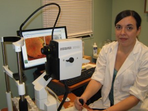
Fundus Imaging
 Fundus photographs are images of the back of the eye. Your doctor may take fundus photographs of your eye to make sure your optic nerve, vitreous (the gel that fills the eye), retina, macula (center of the retina) and blood vessels are healthy.
First, your pupils will be dilated with eye drops. Then you'll look into the special fundus camera. You'll see a series of bright flashes as the camera takes highly magnified and detailed images of your eye. The whole procedure is painless and takes only 5-10 minutes. The photographs may be compared to earlier images taken of your eyes or filed away for future comparison.
Fundus Photography Using a digital fundus camera, images of your eye are taken in color. This can provide your doctor with additional information and documentation of the retina's appearance.
Infrared (IR) Using infrared light instead of typical white light can provide more distinct images of certain portions of the eye. This can be particularly beneficial in patients with developed cataracts.
Red-Free (RF) Red-free photographs filter out the red light and offer a high-contrast image of the vascular system of the eye.
Autofluorescence (AF) Fundus autofluorescence allows for imaging of the retina and identification of retinal diseases that might not be readily apparent. It can determine metabolic changes within the deep retina that are not able to be seen during a routine examination. This approach can be especially helpful to analyze patients with vision loss of undetermined origins or those with a family history of hereditary retinal diseases.
Angiography
Fluorescein Angiography (FA) is a diagnostic test used to evaluate the retina and the retinal blood vessels with the help of a contrast dye (fluorescein). These pictures help doctors evaluate the retina and diagnose and track problems such as diabetic retinopathy, macular degeneration, abnormal blood vessel growth, retinal swelling, retinal detachment, cancer and retinal tumors.
 First, the patient's pupils are dilated with eye drops. Then a few photographs are taken with a special ophthalmic camera. Next, the contrast dye is injected, usually in the patient's arm. The dye travels up to the eye within a few seconds and "lights up" the retinal blood vessels and retinal structures.
Indocyanine Green Angiography (ICG) This is another excellent method for visualizing retinal circulation. In contrast to fluorescein angiography, ICG is of particular value in studying choroidal circulation while investigating macular disease.This test only takes a few minutes to perform.
Optical Coherence Tomography
Optical coherence tomography (OCT) is an advanced technology used to produce cross-sectional images of the retina. These images can help with the detection and treatment of serious eye conditions such as macular holes, macular swelling and optic nerve damage.
OCT uses technology that is similar to CT scans of internal organs, using a scattering of light to rapidly scan the eye to create an accurate cross-section. Unlike other imaging techniques, OCT uses light to produce high resolution images, rather than sound or radiofrequency waves. Your doctor can evaluate and measure each layer of the retina through this image and compare it with normal, healthy images of the retina.
The OCT exam takes about 5 minutes to perform.
Ultrasonography - B-scan
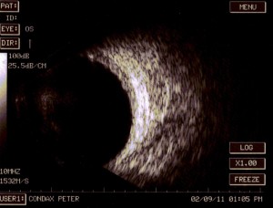
The doctor can perform an ophthalmic ultrasound using simple sound waves that reflect off different tissues in the eye to form an image.
This test is used to determine the extent of a retinal detachment, evaluate the retina and vitreous behind an advanced cataract or hemorrhage and determine the thickness of an intraocular tumor. This test takes a few minutes to perform.